The Amnis ImageStream®X Mark II Imaging Flow Cytometer combines the speed, sensitivity, and phenotyping abilities of flow cytometry with the detailed imagery and functional insight of microscopy. The ImageStream®X MKII systems deliver multi-channel images of every cell in flow, including brightfield, darkfield(SSC) and up to 6 fluorescent markers at high speed. Ultrasmall camera pixel size allows visualization of fluorescence location from the membrane, cytoplasm, subcellular organelles or nucleus at high resolution.The innovative design increases signal to noise ratio providing exceptional photonic sensitivity. A dedicated side scatter laser, adjustable laser intensities, and bright-field imagery for the direct measurement of cell size allow the systems to resolve cell populations more effectively. By overcoming the limitations of both techniques, Imaging Flow Cytometer opens the door to an extensive range of novel applications in life sciences including coolocalization and trafficking, apoptosis and necrosis, autophagy, morphology, etc.
Technical Specifications:
- Objective lens – 60X, 40X, and 20X
- Camera pixel size – 0.1, 0.25, 1 μm2
- Excitation laser – 405 nm, 120 mW; 488 nm 200 mW; 642 nm 150 mW; 785 nm dark field laser
- Six high resolution imaging channels
- INSPIRE and IDEAS software package
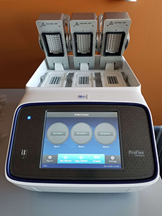
The ProFlex™ Thermal Cycler system combine reliability and performance with the flexible configuration and control features to adapt for specific research needs. Interchangeable block formats allow users to maximize the throughput or run independent experiments concurrently. VeriFlex Blocks provide a better-than-gradient approach to PCR optimization, yielding precise control over three temperature zones.
Simulation Modes
The ProFlex™ PCR system is equipped with Simulation Modes that mimic older thermal cycler's ramp rate. Simulation modes are available for MJ Research PTC 200, Bio-Rad® C1000, Bio-Rad® MyCycler™, Eppendorf® Mastercycler®, Applied Biosystems® 9700, Applied Biosystems® 9600, and Applied Biosystems® Veriti® systems.
VeriFlex™ Blocks: Enhanced PCR Functionality
VeriFlex™ blocks are controlled by multiple Peltier elements (2 separate Peltier elements per 32-well module). This gives users excellent control of the block for precise, "better-than-gradient" optimization experiments, and for running multiple reaction conditions at once for precise incubations and other non-PCR workflows.
Technical Specifications:
| Capacity |
1 x 96-well x 0.2 ml tubes or 3 x 32-well x 0.2 ml tubes |
| Display Interface |
Touchscreen (8.4 in. TFT LCD) |
| Format |
0.2 ml tubes,12-strip wells |
| Peak Block Ramp Rate |
6.0°C⁄sec
|
| Reaction Speed |
Fast, Standard |
| Reaction Volume Range |
10-80 µl |
| Sample Ramp Rate |
± 4.4 °C⁄sec |
| Temperature Accuracy |
±0.25°C (35°C to 99.9°C) |
| Temperature Range (Metric) |
0 to 100.0 °C |
| Temperature Uniformity |
<0.5 °C (20 sec after reaching 95 °C) |
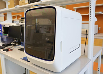
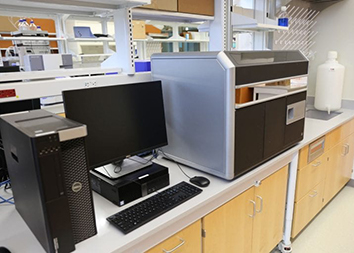
The QuantStudio 7 Flex Real-Time PCR System delivers reliable, sensitive, and accurate quantification with the versatility. It offers optimized protocols, reagents, and intuitive software for the broadest range of quantitative PCR applications. QuantStudio 7 can run hundreds of real-time PCR reactions using TaqMan Array microfluidic cards. Enabling improved well-to-well and instrument-to-instrument data accuracy, the OptiFlex™ System features six decoupled excitation and emission filter channels, with 21 filter combinations for maximum multiplexing and chemistry flexibility.
Key Applications:
- Gene expression
- miRNA profiling
- Long non-coding RNA analysis
- SNP genotyping
- Copy number variation
- Protein thermal shift
- High resolution melt
- Pathogen detection
Technical Specifications:
- Colors: 6 (21 filter combinations)
- Chemistry: SYBR, TaqMan
- Available Blocks: Fast 96-well, 384-well, TaqMan Array Card (384-well microfluidic card)
- Automation: Yes
- Throughput: Medium
- Block change: Block change from front in less than 1 minute, no tools required
- Remote monitoring: Available to monitor up to 15 networked instruments simultaneously
- Intuitive touch screen: Yes

The BD FACSCelesta flow cytometer offers accessible multicolor flow cytometry. The instrument is optimized for working with bright, photo-stable BD Horizon Brilliant dyes, enabling detection of low-density antigens and rare populations. A narrow spectrum of Brilliant dyes minimizes spectral overlap and simplifys experimental design and analysis, allowing simultaneous detection of up to 14 parameters using three lasers. The BD FACSDivaTM software is available to streamline workflow from system setup to data acquisition to data analysis. Together the BD FACSCelesta flow cytometer enables increasingly deep and powerful insights in cell analysis.
Technical Specifications:
- BVR Laser configuration: 405 nm, 488 nm, 640 nm.
| Fluorophores |
Standard Filters |
Standard Mirrors |
|---|
| Blue Laser (488 nm) |
| BD Horizon Brilliant Blue 515, FITC |
530/30 |
505 LP |
| PE |
575/25 |
550 LP |
| BD Horizon PE-CF594 |
610/20 |
600 LP |
| PE-Cy7 or PerCp-CyTM 5.5 |
780/60 or 695/40 |
750 LP or 670 LP |
| Red Laser (640 nm) |
| APC |
670/30 |
N/A |
| BD HorizonTM APC-R700 |
730/45 |
690 LP |
| APC-Cy7 |
780/60 |
750 LP |
| Violet Laser (405 nm) |
| BD Horizon Brilliant Violet 421 |
450/40 |
N/A |
| BD Horizon Brilliant Violet 510 |
525/50 |
505 LP |
| BD Horizon Brilliant Violet 605 |
610/20 |
595 LP |
| BD Horizon Brilliant Violet 650 |
670/30 |
655 LP |
| BD Horizon Brilliant Violet 786 |
780/60 |
750 LP |
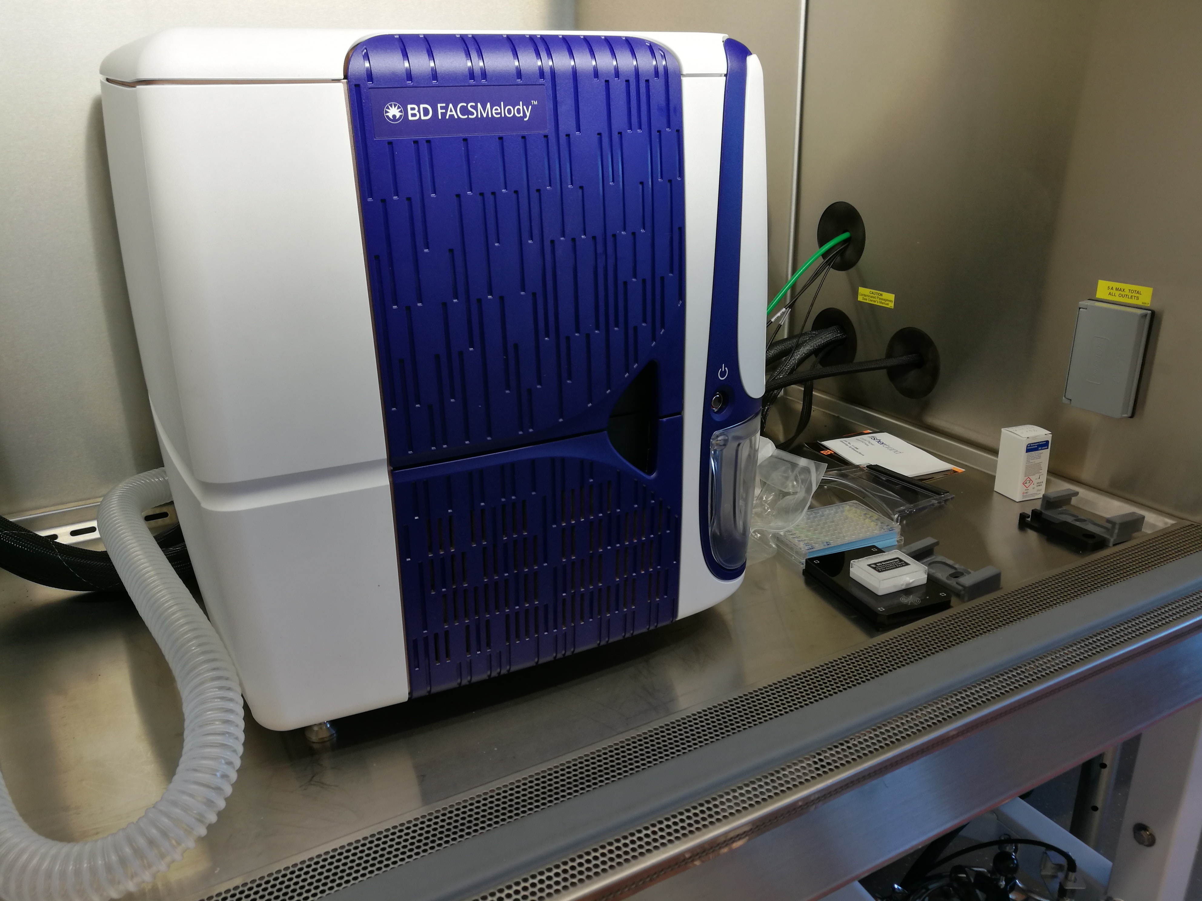
The BD FACS Melody Cell Sorter provides 9 color, 4-way cell sorting with a plate option. The system offers automation technology with BD FACS Chorus™ Software and guides users through the entire cell sorting process with the intuitive interface, on-screen instructions and tips and simple-to-read reports.
Technical Specifications:
• BRV Laser configuration: 405 nm, 488 nm, 640 nm.
• Sort collection devices:5 mL tubes, 96-well plates (using automated stage).
• Sample temperature control
• Aerosol management option (AMO)
• Biological safety cabinet (BSC)
|
Fluorophores
|
Filter
|
Mirror
|
|
Blue Laser (488 nm)
|
|
PE-Cy7
|
783/56
|
752 LP
|
|
PerCP-Cy5.5
|
700/54
|
665 LP
|
|
PE, PI
|
586/42
|
560 LP
|
|
FTIC, GFP, Alexa 488
|
527/32
|
506 LP
|
|
Red Laser (640 nm)
|
|
APC-Cy7
|
783/56
|
752 LP
|
|
Alexa 700
|
720/30
|
705 LP
|
|
APC, Alexa 647
|
660/10
|
660/10
|
|
Violet Laser (405 nm)
|
|
BD Horizon Brilliant Violet 786
|
None
|
755 LP
|
|
BD Horizon Brilliant Violet 510
|
528/45
|
500 LP
|
|
BD Horizon Brilliant Violet 421, DAPI
|
None
|
448/45
|
The BD Rhapsody Single-Cell Analysis System allows high-throughput capture of multi-omic information from single cells using a simple cartridge workflow and a multitier barcoding system. The resulting captured information can be used to generate various types of next-generation sequencing (NGS) libraries. 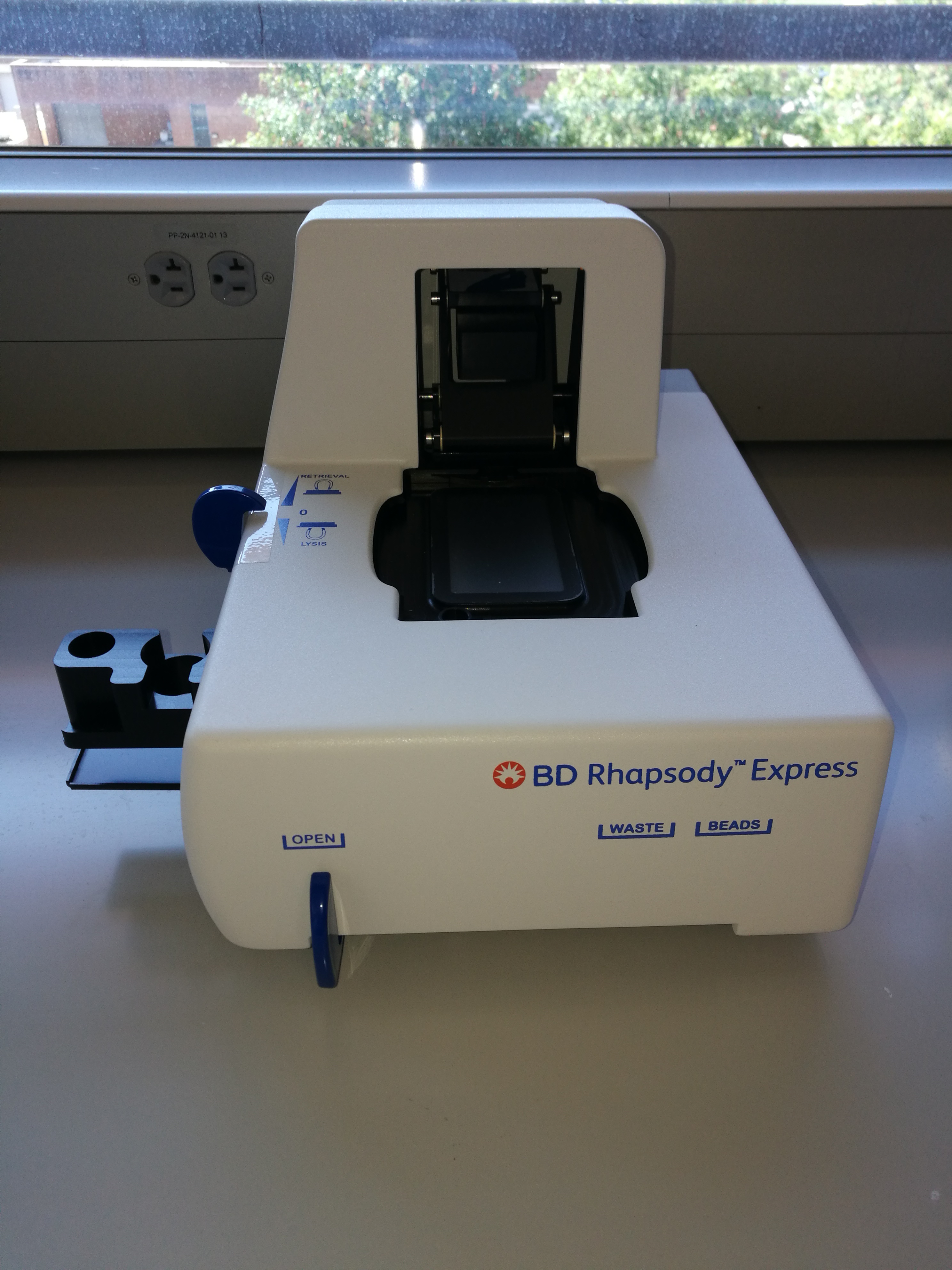
Features:
- Capture and analyze fragile cells: Granulocytes, neutrophils, CAR-T cells, stem cells, tumor xenograft-derived cells, myeloma, T cells, NK cells and more
- Up to 80% cartridge capture rate
- Broad range of cell: 100 - 40,000 cells per cartridge
- Microwell technology and no sample loss due to clogging of channels
- High correlation with flow data
- Subsample beads and Archive beads
- Minimal batch effects
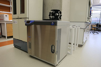 The FreeZone 6 L Console Freeze Dryer is designed for lyophilizing moderate to large sample loads or numerous small container batches. The system is equipped with a 12-port chamber. Two sizes of Fast Freeze flasks are available 600 mL and 900 mL. Interior PTFE coating provides corrosion resistance.
The NanoDrop 2000c is a full-spectrum UV-Vis spectrophotometer equipped with both a sample-retention system for microvolume samples and cuvette measurment. Simplified software displays a full-spectrum and tabulates results for easy data interpretation.
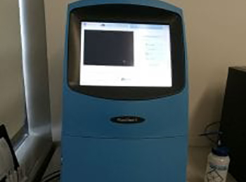 The FluorChem E from ProteinSimple features simplied touch screen control, straightforward workflow for detection of chemiluminescent, colorimetric and UV fluorescent gels and blots. A 8.3 megapixel, high-resolution CCD camera with 5-log dynamic range enables capturing both faint and bright bands in single expsoure.
The Seahorse XFe96 Analyzer simultaneously measures the two major energy pathways of the cell — mitochondrial respiration and glycolysis — in live cells using a label-free, solid-state sensor cartridges in a specialized 96-well format. The Seahorse XFe96 Analyzer works with all cell types, including primary cells, cell lines, suspension cells, as well as islets, C. elegans, yeast, and isolated mitochondria.
Technical Specifications: 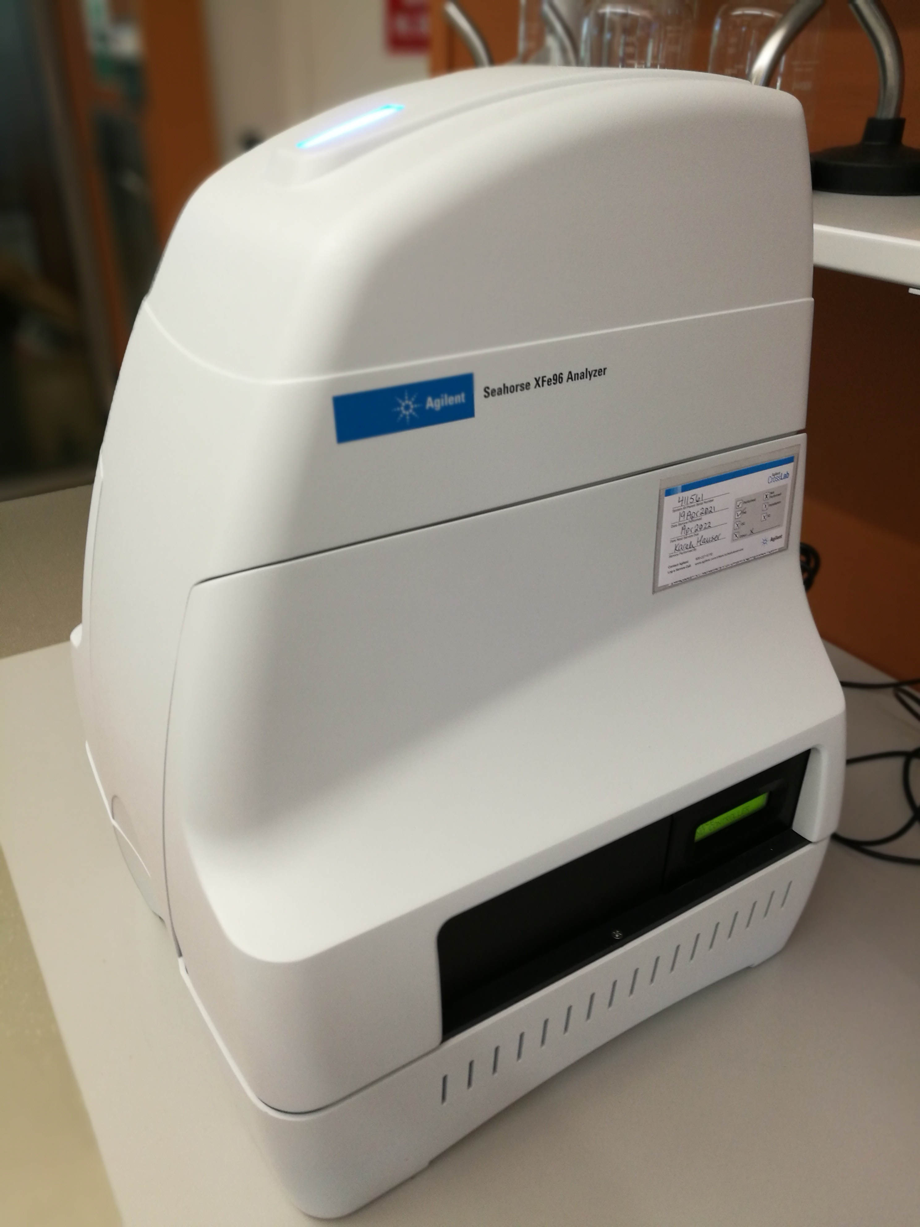
• Primary Measurements Made: OCR & ECAR
• Assay Format: Live cells in a specialized 96-well microplate
• Injection Ports: 4 per well, 25 uL each
• Assay Running Volume: 80-200 uL/well
• Sample Requirement: 4,000-500,000 cells/well
Key Features:
• Compatibility with adherent and suspension cells as well as isolated mitochondria and non mammalian samples
• Ability to perform up to four independent injections per well with automatic mixing • Automatic calculation of oxygen consumption rate (OCR) and extracellular acidification rate (ECAR)
• Simultaneous measurement of OCR and ECAR in the same well
• Sensitivity for small sample sizes
• Label-free detection in live cells, in real time
• Windows-compatible desktop analysis software (Wave) for plotting, reporting, analyzing, and exporting your Agilent Seahorse XF data

The Tecan Spark microplate reader offers Absorbance Mode, Top and Bottom Fluorescence Mode, Luminescence Mode, Fluorescence Polarization Mode, and Brightfield Imaging. This multimode system is optimized for a wide range of life science research applications:
- ELISA
- Low-volume DNA/RNA quantification
- Nucleic acid labeling efficiency
- Protein quantification
- Reportergene assays
- Cell counting and viability
- Confluence assessments
- Cell migration and wound healing
Technical Specifications:
- Absorbance Mode
- Top and Bottom Fluorescence Mode
- Luminescence Mode
- Fluorescence Polarization Mode
- Brightfield Imaging for Cell Counting and Confluence
- Temperature control (18 C-42 C)
- CO2 and O2 Control
- Cuvette Port
- Lid lifter plus Humidity Control Cartridges
- Nanoquant Plate for Microvolume Nucleic Acid Quantification
- Magellan Data Analysis Software
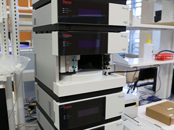
Thermo Scientific DionexTM UltiMateTM 3000 HPLC (UHPLC) system meets everyday HPLC needs while fully supporting UHPLC applications. The system is designed for pressures up to 620 bar (9,000 psi) at flow rates of up to 10 mL/min, detector data collection rates up to 100 Hz, and injection cycle times as low as 15 s. SmartFlowTM and SpinFlowTM ensure optimal flow and gradient performance. Detection versatility is achieved by one highly sensitive Fluorescence Detector and one low-noise, high-linearity Variable Wavelength Detector. Chromeleon 7 Chromatography Data System software guides user from samples to results through streamlined chromatography workflows.
Technical Specifications:
- Thermo Fisher UltiMate 3000 HPLC System
- SRD-3400 Solvent Rack with integrated analytical 4-channel
- LPG-3400SD Low Pressure Mixing Gradient Pump
- WPS-3000TSL Analytical Autosampler
- TCC-3000SD Thermostatted Column Compartment
- FLD-3400RS Fluorescence Detector
- Analytical Flow Cell for FLD-3000 Series, SST, 8 μL Volume
- VWD-3400RS Rapid Separation Variable Wavelength Detector
- Analytical Flow Cell for VWD-3000 Series, SST, 11 μL Volume
- AFC-3000 Automated Fraction Collector
- Chromeleon 7 Chromatography Data System
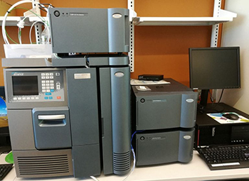
Waters Gel Permeation Chromatography (GPC) system allows accurate measurement of size distribution on polymer samples. The GPC is coupled to Alliance HPLC system, which features integrated fluidics providing a low-dispersion design. Up to four eluents can be automatically conditioned and blended for isocratic, dial-a-mix, AutoBlend, TM or gradient operation in order to minimize user error and simplify method transfer. The detection module contains three highly sensitive detection choices including a differential refractive index detector, a UV/Vis detector, and a photodiode array detector. In addition, Waters Informatics and data handling solutions enables efficient, streamlined results management.
Technical Specifications:
- Alliance HPLC Systems
- e2695 XE Separations Module w/CH (Blue)
- Automatically process up to 120 samples from 5 x 24 carousels
- Sample heating/cooling and column heating capabilities
- Optimized for routine LC and LC/MS analyses
- 2414 Refractometer Detector (blue)
- Differential refractive index detectors for detecting components without UV absorption
- With a wide linear dynamic range for quantitative methods
- Thermally isolated optics bench for temperature stability and performance reproducibility
- 2489 UV/Visible Detector
- Sensitive and versatile dual-wavelength absorbance detector
- Enhanced sensitivity via patented TaperSlit” flow cell
- Low noise performance (<5 μAU)
- Lamp optimization for S/N performance over the lifetime of a Deuterium lamp
- 2998 Photodiode Array Detector
- Trace impurity detection and quantitation in conjunction with spectral analysis
- Enhanced sensitivity via patented TaperSlit” flow cell
- Low noise performance of <10 μAU
- Simultaneous quantification of high- and low-level components
- Empower 3 System Software with GPC/SEC Option
 Bruker VERTEX 70 FT-IR spectrometer is designed to be versatile for demanding applications in the analytical and research laboratory. All optics are gold coated for high optical throughput. The standard spectral range covers 8,000 to 350cm-1. Range extension packages are available to extend the measurement region from the far- to the near-IR and the visible regions (28,000-15cm-1). Standard resolution is 0.4cm-1 with an option for 0.16cm-1. A scan rate of > 15 spectra/sec at 8cm-1 is standard and can be upgraded to higher rates of 58 spectra/sec at 16cm-1. The Step-Scan technique is also available for kinetic experiments in the µs and ns range. VERTEX 70 is equipped with wide band spectral range extension VERTEX FM for mid-IR and far-IR. Covers spectral range from 6.000-80cm-1, optionally extendable up to 9,200cm-1, suitable for spectral resolution up to 0.4cm-1, reduced signal to noise ratio at 610cm-1. The Platinum-ATR-accessory, with the robust diamond crystal, enables a fast and reliable FT-IR-analysis of solids and liquids, without sample preparation. The ergonomic one-finger clamp mechanism permits a comfortable and fast analysis of solids.
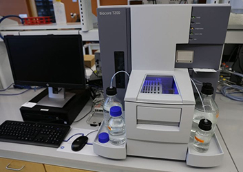 GE Healthcare Biacore T200 system offers label-free, versatile assays for studying biomolecular interactions. The system provides precise quantification of kinetic, affinity, concentration, specificity, selectivity, comparability, and thermodynamic interaction data. Wizard templates and customizable methods/evaluation tools are available for both common assays and sample specific applications. The system’s extreme high sensitivity enables affinity analysis of the smallest organic compounds, interaction study of rare or sensitive targets, and measurement of kinetic constants over a broad range.
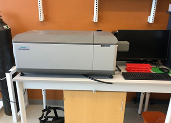 In addition to the standard CD, LD, and absorbance scanning, the JASCO J-1500 Circular Dichroism Spectropolarimeter offers up to four simultaneous measurement modes with a wide range of sampling accessories. These accessories enable measurements of a variety of samples, from liquids to films to solid-states. Peltier temperature control system is coupled with six position turreted to run thermal melts, providing researchers with CD and thermodynamic data sets for conformational and folding studies. The extended wavelength range allows measurements to be obtained in both the vacuum-UV and NIR spectral regions using the standard PMT detector (163 – 950 nm).
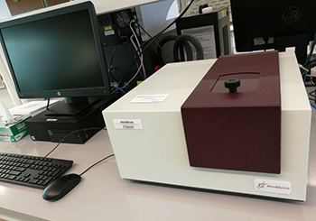 The new NanoBrook Omni particle size and zeta potential analyzer provides fast, routine, submicron measurements of size and zeta potential. Most measurements only take a minute or two based on the principles of Dynamic Light Scattering (DLS) for particle sizing and distribution, and on Doppler velocimetry (electrophoretic lightscattering, ELS) for zeta potential. The instrument also includes Phase Analysis Light Scattering (PALS) measurements for samples with low mobilities (saline, PBS).
 The qNano Gold measures particles using the Tunable Resistive Sensing (TRPS) principle. It measures individual particles with the highest accuracy and repeatability available for, particle size and real distribution, concentration and surface charge.
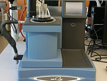 A research quality Dynamic Mechanical Analyzer (DMA) that provides viscoelastic measurements on materials from 1000 Pa to 1000 GPa. The temperature and frequency range of operation are -150 to 600 º C and 0.01 to 200 Hz, respectively. The force range is 0.0001 to 18 Newtons. The amplitude range is 0.5 to 10,000 microns with resolution to 1 nanometer over the entire 25mm of drive shaft travel using a linear optical encoder. The Q800 provides sensitive force measurements down to 0.0001 Newtons, which is ideal for films, fibers and other materials with low stiffness. The module is designed with a full VGA touch control color screen, automated furnace movement, and easy clamping accessibility. The Q800 also includes Advantage ™ software for complete experimental control of all Q Series ™ modules. It offers extensive setup templates and wizards, on-line help menus with diagnostics and multimedia support, plus Universal Analysis 2000, the most comprehensive data analysis program available. Other features include real time data analysis, autoanalysis, ability to create up to 20 simultaneous curve overlay plots, a customer report generator using Microsoft Word and Excel templates, spreadsheet text files, PCX and HPGL graphics files, and ability to export ASCII data files.
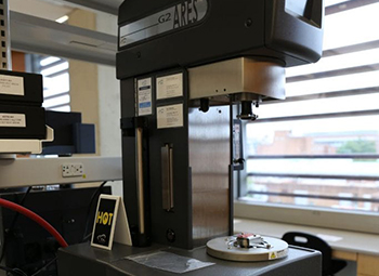 To achieve excellent accuracy in viscosity and viscoelastic material characterization, the TA ARES-G2 Rheometer offers decoupled stress and strain measurements free of instrument artifacts over wide ranges of stress, strain, and frequency. The system features separate motor and transducer technology, independent key component control, and intuitive TRIOS software. Normal Force Rebalance Transducer (FRT) module provides wide torque range and normal force range. Brushless DC motor features air bearings for smooth friction free motion, a high-resolution optical encoder for oscillatory angular displacements down to 1 μrad, and unlimited strain range in steady testing. Motor provides a wide range of angular velocities, and angular frequencies. The TA ARES-G2 is the most advanced rotational rheometer for research and material development.
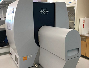 The Bruker SkyScan 1276 desktop X-Ray Microtomograph is designed for fast, in vivo scanning of small lab animals and biological samples. The large field of view (up to 80 mm wide and more than 300 mm long) allows full body mouse and rat scanning. High spatial resolution down to 6 μm voxel size and large image size are achieved by combining continuously variable magnification and 11 Mp X-ray CCD camera (2.8 μm pixel size). Other features include round and spiral scanning enabled by continuous gantry rotation, low dose imaging allowing longitudinal preclinical studies, and fast GPU-based 3D reconstruction from cross sections.
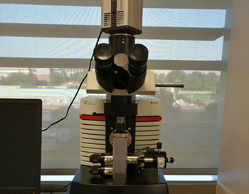 The Low Voltage Electron Microscope 5 is designed for a broad range of applications in material sciences, such as nanomaterials, polymers and biomaterials, and in life sciences, such as drug discovery and delivery, pathology and virology. The LVEM5 offers a high throughput benchtop solution with nanometer resolutions. The LVEM5 is approximately 90 % smaller than classical electron microscope. There is no need for dark room or cooling water. The LVEM5 provides nanometer level resolution in TEM (Diffraction included) and SEM. Low accelerating voltage (5 kV) results in increased electron scattering and enhanced contrast on biological, organic and light materials. Coupled with TEM BOOST module, a hardware-based enhancement of TEM imaging mode, LVEM 5 provides increased total magnification and higher resolving power in the TEM images.
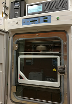 Sartorius Incucyte S3 Live-Cell Analysis Instrument offers options of tracking and monitoring cells for
 The NanoWizard 4a BioScience AFM offers atomic resolution and fast scanning (up to 100Hz) with scan range of 100μm. JPK’s unique design enables imaging samples in liquids under physiological conditions. With highest mechanical/thermal stability on an inverted microscope and lowest noise level for all AFM components, NanoWizard 4a is optimized for long term experiments even on living cells. The improved QI mode delivers quantitative imaging with the highest resolution for single molecules, live cells or tissues. NanoWizard 4a comes with a wide range of accessories and modes providing full flexibility for any application.
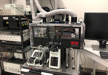 The FLUOVIEW FV3000 laser scanning confocal system offers high sensitivity and speed required for 6D (x,y,z,t,λ,p) live cell and tissue imaging. FV3000 supports complete workflows from live cell imaging through image processing, like deconvolution, and segmentation. The system features a new spectral detection concept for true multichannel spectral imaging with high sensitivity detection in multiple dynamic ranges. The customized 730 nm diode laser and near IR GaAs detector further extends the detection range. With PicoQuant turn-key upgrade kit, time-resolved techniques (e.g. FLIM) can be performed in 2 channels (440 and 485 nm).
 The Leica Biosystems Suite offers histology and immunohistochemistry at light microscopic level. Automated systems are available for tissue processing, embedding, and staining: